

What is Myxomatous Mitral Valve Disease?
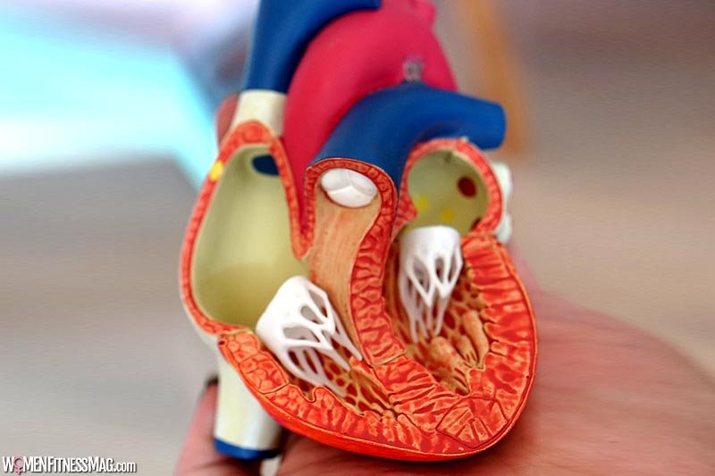
Myxomatous mitral valve disease refers to a degenerative condition wherein there is improper closure of the mitral valve of the heart. This is due to the thickening of the valve forming small nodules on the edges of the leaflets preventing the tight closure of the valves. The mitral valve sits between the left ventricle and the left atrium of the heart.
In myxomatous mitral valve disease, the leaflet of the mitral valve sticks out into the left atrium during the contraction of the heart. It is the mitral valve that keeps the forward direction of the flow of blood. But in the case of myxomatous mitral valve disease, due to the improper closing of the mitral valve, the backward direction of blood flow into the left atrium happens – mitral regurgitation.
Myxomatous mitral valve disease is also called in several terms like Mitral Valve Prolapse or Click-murmur Syndrome or Floppy-valve Syndrome or Billowing Mitral Valve Syndrome or Barlow’s Syndrome.
Myxomatous mitral valve disease is typically present in people with rheumatic heart disease, Marfan’s syndrome, or Graves’ disease. Treatment is not necessary in most cases of myxomatous mitral valve disease. However, for some life-threatening cases, medications and surgeries are recommended.
Signs and Symptoms
Although myxomatous mitral valve disease is usually a lifelong disorder, many people with this condition are asymptomatic, they do not experience any symptoms. Only when diagnosed, people will be surprised to learn that they have a heart disorder.
When blood is leaking backward through the valve, signs, and symptoms may occur. In myxomatous mitral valve disorder, symptoms may vary widely from person to person. Symptoms tend to be mild and may eventually develop gradually. However, serious symptoms of myxomatous mitral valve disease tend to occur most often in men older than 50. Some patients may exhibit symptoms which may include:
- Anxiety
- Dizziness
- Chest pains
- Syncope or fainting
- Lightheadedness
- Fatigue
- Panic attacks
- Arrhythmia or irregular heartbeat
- Migraine – one-sided headache
- Dyspnea or shortness of breath
- Difficulty in breathing when lying flat or during physical activity
- Orthostatic hypotension – low blood pressure in lying position
Risk Factors
There are few health conditions that may contribute to the increase in the person’s risk of developing myxomatous mitral valve disease. This disorder may develop in any given age of a person. Myxomatous mitral valve disease may run in families and may be linked to several other conditions, such as:
- Graves’ Disease
- Muscular Dystrophy
- Marfan Syndrome
- Ebstein’s Anomaly
- Scoliosis
- Ehlers-Danlos Syndrome
Causes
The major cause of the prolapse of the myxomatous mitral valve is the degeneration of valvular tissue that causes the leaflets of the valve to enlarge and stretch. This condition will result in a bulge into the left atrium that will prevent the valve from closing properly.
Aside from tissue degeneration, functional myxomatous mitral valve prolapse may also appear due to conditions such as:
- Dilated cardiomyopathy
- Myocardial ischemia
- Hypertrophic cardiomyopathy
Diagnosis
In examining the myxomatous mitral valve prolapse, initially, the doctor will hear the heart sounds of the person to rule out any abnormalities in the heart sounds like clicking sound which is indicative of myxomatous mitral valve disease. It also permits the doctor to check on the presence of mitral valve regurgitation.
Other tests to diagnose Myxomatous Mitral Valve disease may include:
- Stress Test
A stress test is conducted to check the presence of mitral valve regurgitation, which restrains the ability of the patient to exercise. In the stress test, the patient is required to exercise or to take medications under the supervision of a doctor to increase the pulse rate and make the heart work more. A stress test is a powerful tool in assessing the presence of coronary heart disease. - Chest X-ray
Since a chest X-ray can provide the image of the vital organs in the body such as lungs, heart, and also the blood vessels, the doctor may require the patient to have this done to detect any heart enlargements. - Electrocardiogram (ECG)
The electrocardiogram is a non-invasive diagnostic test that uses electrodes in determining the muscular and electrical activities of the heart. The result of the electrical signals recorded will help the doctor determine any defects in the structure and rhythm of the heart. - Echocardiogram
An echocardiogram is a diagnostic procedure that is used to evaluate the heart. The echocardiogram procedure, high-frequency sound waves are used to create images of the structures of the heart of the patient. It may also help in the evaluation of the blood flow through the mitral valve and the amount of blood leakage or regurgitation if present. - Coronary Angiogram
Coronary Angiogram is a specialized type of X-ray technique used for the examination of the coronary artery. This is not a specific diagnostic tool for myxomatous mitral valve prolapse test. However, a coronary angiogram can also help detect its presence after it is done in the examination of other heart conditions.
Treatment
For some cases, the doctor may prescribe medications for the patient who displays mitral valve regurgitation. But in case the patient is not responsive to the prescribed medications, surgery may be recommended, and as follows:
- Medications
For some that developed the symptoms of mitral valve regurgitation, the doctor may recommend the individual to take some medications. The common medications being prescribed include aspirin, diuretics, beta-blockers, anti-arrhythmic, and some blood thinners. Long-term use of these medications could lead to mental anxiety in some patients. - Surgery
With or without evident symptoms, the surgery is the most preferred option in treating patients with mitral valve regurgitations.There are two approaches to surgery as an option of treatment:
- Valve Repair
In most cases of mitral valve regurgitations, valve repair is the most preferred approach. The mitral valve consists of two flaps and a ring-like structure called mitral annulus. The mitral annulus constitutes the anatomical connection between the left ventricle and the left atrium and serves an insertion site for the leaflet tissue to the heart muscle.To repair the valve, the excess mass is removed or the leaflets of the valve are reconnected to facilitate the tight closing of the leaflets. There are cases wherein the valve is also repaired by reinforcing or replacing the annulus.
- Valve Replacement
For some patients where repair of valves is not a doable option of treatment, valve replacement is done. As the term implies, it involves the replacement of the defective valve with an artificial or prosthetic valve.There are two types of artificial valves: mechanical valves and tissue valves – made from animal tissues.
Mechanical valves are the ones that can last longer but the individual will be advised to take anticoagulant medications throughout their life.
Whereas, the tissue valves may not last very long and wear out more easily. And for those who will prefer to have the tissue type of valve for the replacement, taking anticoagulant medications is not necessary.
Related Videos about Myxomatous Mitral Valve Disease :
Mitral Valve Disease
Mitral valve disease (regurgitation, stenosis) – causes, symptoms & pathology
https://www.youtube.com/watch?v=jYzjKOWXVww
Heart Valve Disease in Women
Myxomatous Mitral Valve Disease
pathophysiology of myxomatous mitral valve disease, myxomatous mitral valve icd-10, acvim myxomatous mitral valve disease, myxomatous tricuspid valve, mitral valve prolapse and coronavirus, mitral valve prolapse echo, mitral valve prolapse treatment, what should i avoid if i have mitral valve prolapse,