

Dermoscopy is an examination of skin lesion through a handheld instrument called dermatoscope. The procedure uses a transilluminating light and standard magnifying optics to scan below skin surface which can be magnified tenfold.
Dermoscopy is a noninvasive medical procedure which is commonly used by medical practitioners to examine several types of skin conditions. Basically, the medical procedure allows the medical practitioner to identify benign from malignant tumours, such as in cases of the diagnosis of melanoma and other cancerous lesions which include basal cell carcinomas, angiomas, cylindromas, seborrheic keratosis, dermatofibromas, and squamous cell carcinomas.
The medical examinations require the use of dermatoscope, allowing physicians in determining the surgical margin for skin cancers that are difficult to define. If you’re in the market for dermatoscopes online, find a trusted medical supplier to ensure you get quality equipment you can trust.
Historically speaking, dermoscopy is utilised in order to aid dermatologists from identifying skin conditions and other lesion details which a physician cannot examine with their naked eye. Today, digital dermoscopy is now available, helping many clinicians in monitoring potentially cancerous skin lesions. In this procedure, digital images are processed, gathered and stored which will be compared afterwards from the patient’s next medical visit.
How Does Dermoscopy Work?
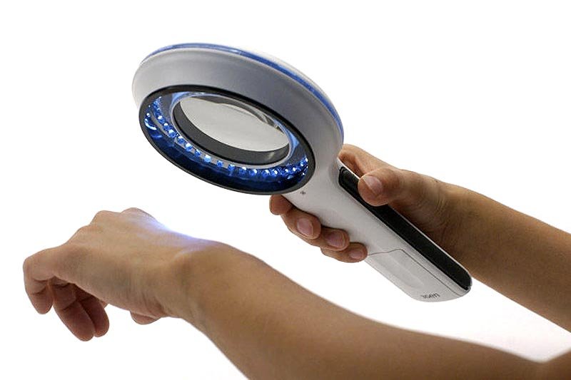
As earlier discussed, dermoscopy used a handheld microscope tool that your clinicians holds up to the affected skin area to examine them closely. Basically, the dermatoscope combines high light intensity and high magnification, allowing your clinician to see up close the diagnostic features of your lesion that cannot be seen through the naked eye. In fact, scientific studies provide that these medical techniques have shown to be more accurate when it comes to diagnosing cancerous moles than the naked eye alone. The procedure is known to get rid of unnecessary procedures and can easily help a clinician in determining if a skin spot is needed to be biopsied.
If you are advised to undergo this type of procedure, your physician will certainly give you a few information regarding the process. Basically, the whole medical procedure is very simple and no strict regimen is needed before the actual day of the examination.
After doing the initial medical checkup and after acquiring your overall medical condition results, confirming the necessity of undergoing dermoscopy, your physician will inform you of the findings so you could make the necessary preparation for it.
Despite the simplicity of the whole medical procedure, the medical expertise of a medical practitioner is still needed to make sure that the results are accurate. In doing the procedure, your clinician will apply an appropriate amount of liquid alcohol or gel on the pigmented skin area; the gel used in an ultrasound scanning can also be used in this medical procedure. The application of liquid gel or alcohol is imperative in order to enhance the clearness of the stratum corneum, allowing a transparent visualisation of specific structures of your epidermis, your papillary dermis and your dermo-epidermal junction.
Once the liquid gel has been applied on your skin, your clinician will place the dermatoscope on the top of the lesion and gently press against the pigmented skin area. Your clinician will eventually add more pressure as he/she presses the medical device in order to completely eliminate air bubbles. As of now, there are three types of dermoscopic techniques used by clinicians in conducting the medical procedure which includes :
- polarized contact
- polarized non-contact
- classic or standard contact.
To put it briefly, polarized dermoscopy gives more advanced reactivity for diagnosing skin cancer due to its particularization to improve crystalline and vascular structures while non-polarized dermoscopy enhances particularity as it allows better inspection of other structures commonly detected in benign lesions. Objectively, both polarized and nonpolarized dermoscopy provide complementary information, aiding many medical experts in ascertaining whether a biopsy is still needed.
Conclusion
Advanced melanoma cannot be remedied by treatment. However, several studies provide that early detection of cancer at its initial stage shows a significant chance for its recovery. Hence, a medical procedure that provides some relief, such as dermoscopy, to help alleviate the suffering of patient battling melanoma offers comfort and hope not only to the patient but to the family and the entire medical industry as well.
Indeed, beyond it noninvasive nature, dermoscopy provides a wide array of medical benefits both to the patients and medical practitioners alike as it extends not only solution but also gives solace to those who are suffering from severe skin diseases.
Related Videos about What is Dermoscopy and How Does It Work?
What is a dermoscopy?
Accuracy of Dermoscopy
https://www.youtube.com/watch?v=6VJKlNytemA
Watch & Learn: Dermoscopy
Dermoscopy in Practice
https://www.youtube.com/watch?v=bXoChrp4Ss0